Fetal ultrasound is a test done during pregnancy that uses reflected sound waves to produce a picture of a fetus camera.gif, the organ that 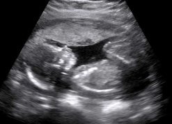 nourishes the fetus (placenta), and the liquid that surrounds the fetus (amniotic fluid). The picture is displayed on a TV screen and may be in black and white or in color. The pictures are also called a sonogram, echogram, or scan, and they may be saved as part of your baby’s record.
Fetal ultrasound is the safest way to check for problems and get information about your fetus, such as its size and position. It does not use X-rays or other types of radiation that may harm your fetus. It can be done as early as the 5th week of pregnancy. The sex of your fetus can sometimes be determined by about the 18th week of pregnancy.
Pregnancy: Should I Have an Early Fetal Ultrasound?
A combination of screening tests using ultrasound may be done in the first trimester to look for birth defects, such as Down syndrome. The first-trimester screening test uses an ultrasound measurement of the thickness of the skin at the back of the baby’s neck (nuchal translucency) and the blood levels of free beta-HCG and a protein called pregnancy-associated plasma protein A (PAPP-A) to check for problems.